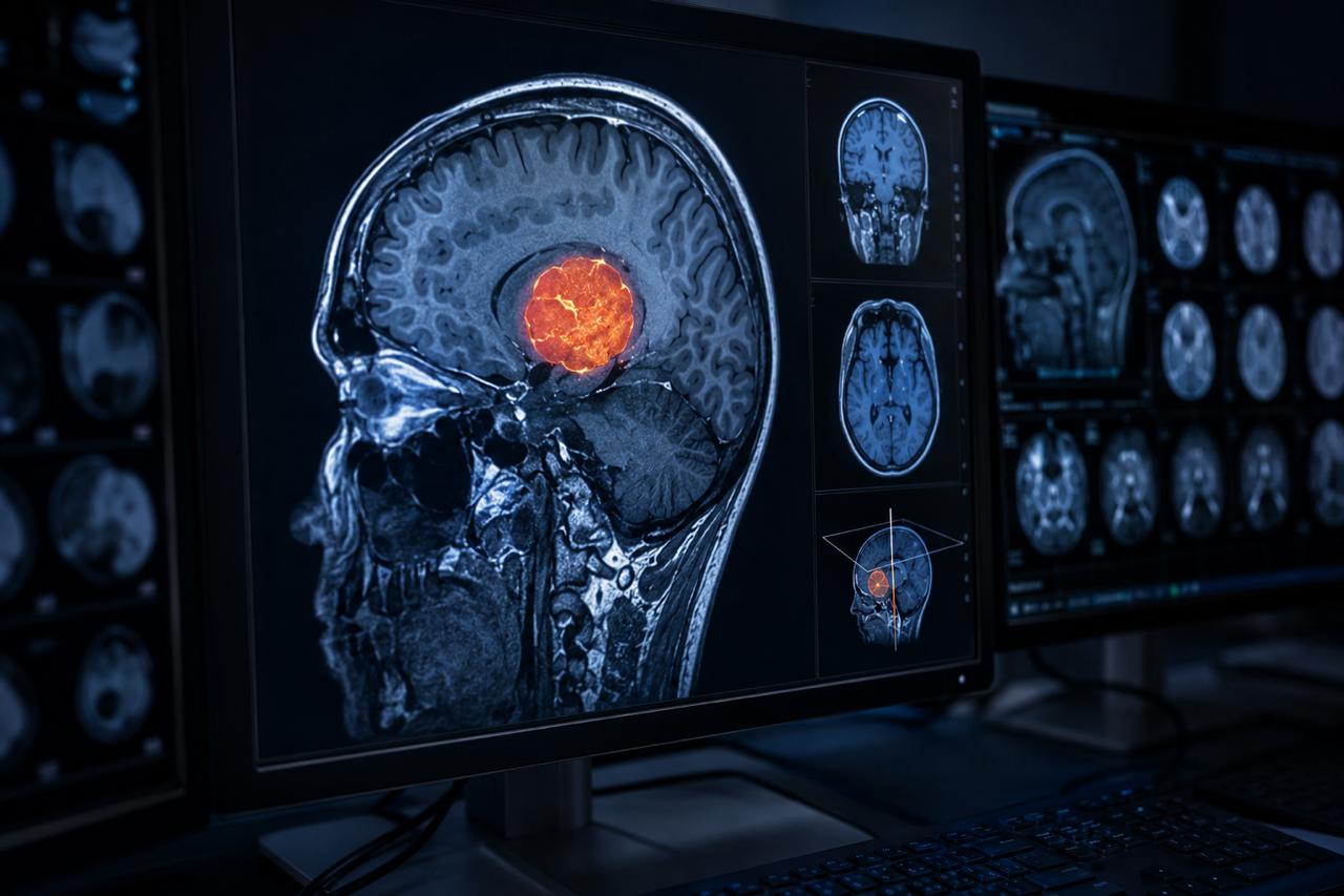
Turning patient imaging into surgical intelligence — the clinical foundation for every case we work on.
Before a surgeon can plan, they need to see. Not flat slices of a standard DICOM viewer — but a precise, three-dimensional reconstruction of the patient's anatomy that reflects the reality of what they will encounter in theatre.
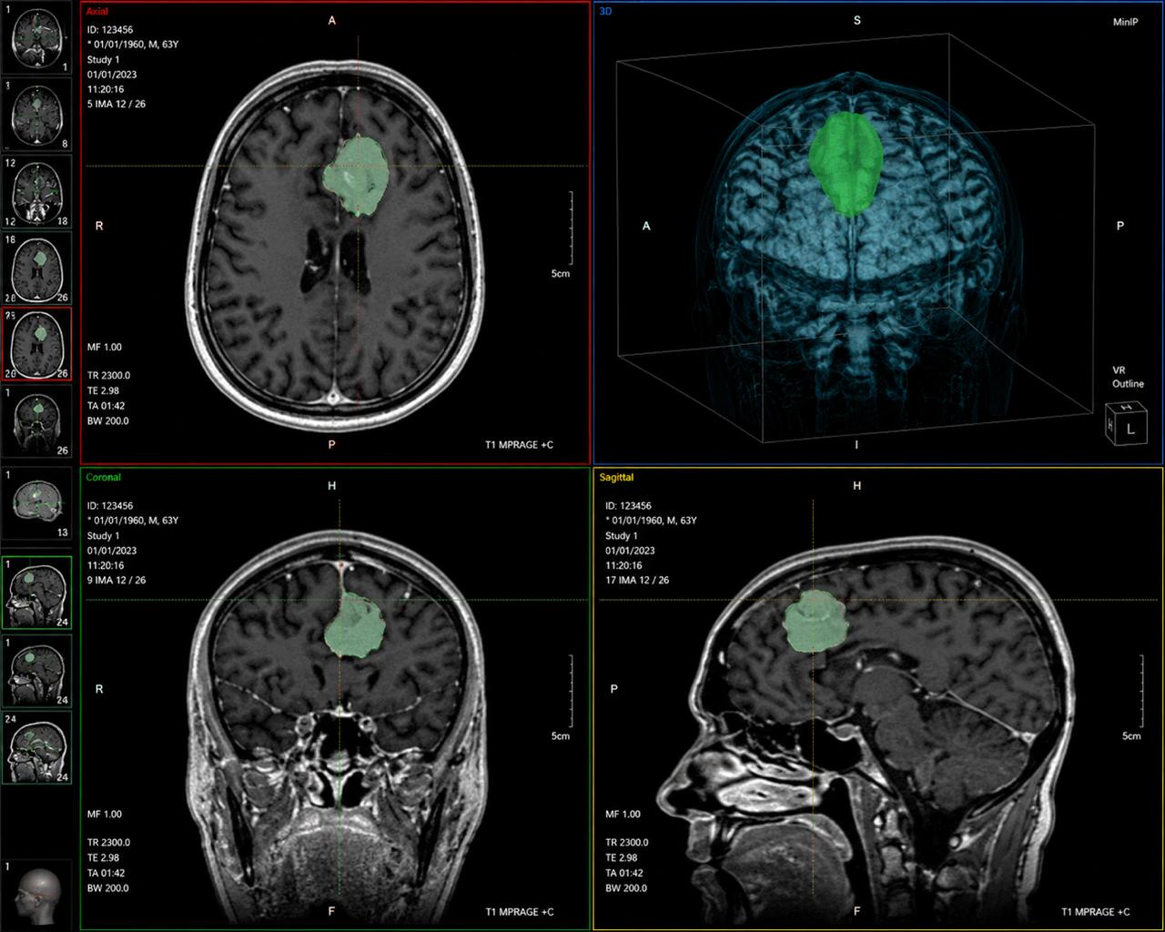
We process CT and MRI imaging data from your institution's existing scans — no additional imaging required. Our clinical team segments each anatomical structure with precision, producing accurate 3D reconstructions in a format meaningful for surgical planning.
Four core deliverables — each designed to give the surgical team exactly what they need before entering the theatre.
Full patient-specific model segmented from CT or MRI DICOM data. Every surface reproduced with clinical accuracy — bone, soft tissue, vasculature, and pathological structures clearly delineated.
Tumour, lesion, or abnormal structure isolated and rendered distinctly from surrounding anatomy. Critical neurovascular relationships mapped and visualised with surgical-grade precision.
A structured walkthrough with the surgical team to review the reconstruction, discuss anatomical findings, map the surgical approach, and define the operative strategy before theatre.
STL and surgical-compatible file formats delivered for downstream use in implant design, guide manufacturing, or anatomy model production. Full file traceability included.
| Output | Detail | Turnaround |
|---|---|---|
| 3D Anatomical Reconstruction | Full segmented model from CT or MRI DICOM — bone, soft tissue, vasculature, pathology | 3–5 working days |
| Pathology Delineation | Tumour or lesion isolated and rendered with neurovascular mapping | 3–5 working days |
| Digital Review Session | Structured walkthrough with surgical team — anatomy, approach, strategy | Scheduled with team |
| STL / Surgical File | Compatible with implant design, guide manufacturing, and anatomy model workflows | Included with model |
| Urgent Cases | Priority processing for time-sensitive surgical cases — contact us directly | 48 hours |
Complex spinal and craniovertebral junction surgery, skull base and posterior fossa tumour resection, paediatric cases with developmental anatomical variation.
Reconstruction with compromised or atypical anatomy, revision arthroplasty planning, deformity correction cases requiring precise preoperative understanding of bony geometry.
Osteotomy and reconstruction planning, orbital anatomy mapping, mandibular defect assessment, and midface reconstruction planning requiring millimetre-level accuracy.
Bone and soft tissue tumour resection planning, margin definition, and MDT pre-surgical briefing for cases where resection geometry determines reconstruction design.
Vascular structure reconstruction and cardiac anatomy modelling for complex access planning, device selection, and surgical rehearsal.
Scaled anatomical reconstructions for developmental anomalies and rare paediatric anatomies where standard surgical reference points do not apply.
Submit your case with DICOM imaging data and clinical brief. We confirm receipt and assign a clinical lead within 24 hours.
DICOM files segmented structure by structure. Every surface validated against clinical benchmarks before reconstruction is finalised.
Reconstruction shared with the surgical team. We walk through the anatomy together and incorporate all clinical feedback.
Final segmented model and surgical files delivered with full documentation. Ready for downstream use in implant design or guide production.
Our clinical team remains available for queries through to the day of surgery and beyond.
Custom implants in titanium, PEEK, and PMMA — engineered from individual anatomy, surgeon-reviewed before manufacture.
Learn more →Precision cutting, drilling, and positioning guides that translate the preoperative plan to the surgical field accurately.
Learn more →High-fidelity physical replicas for surgical rehearsal, MDT briefings, and patient consent at true scale.
Learn more →Submit your case and our clinical team will be in touch within 24 hours to discuss your specific requirements and imaging data.